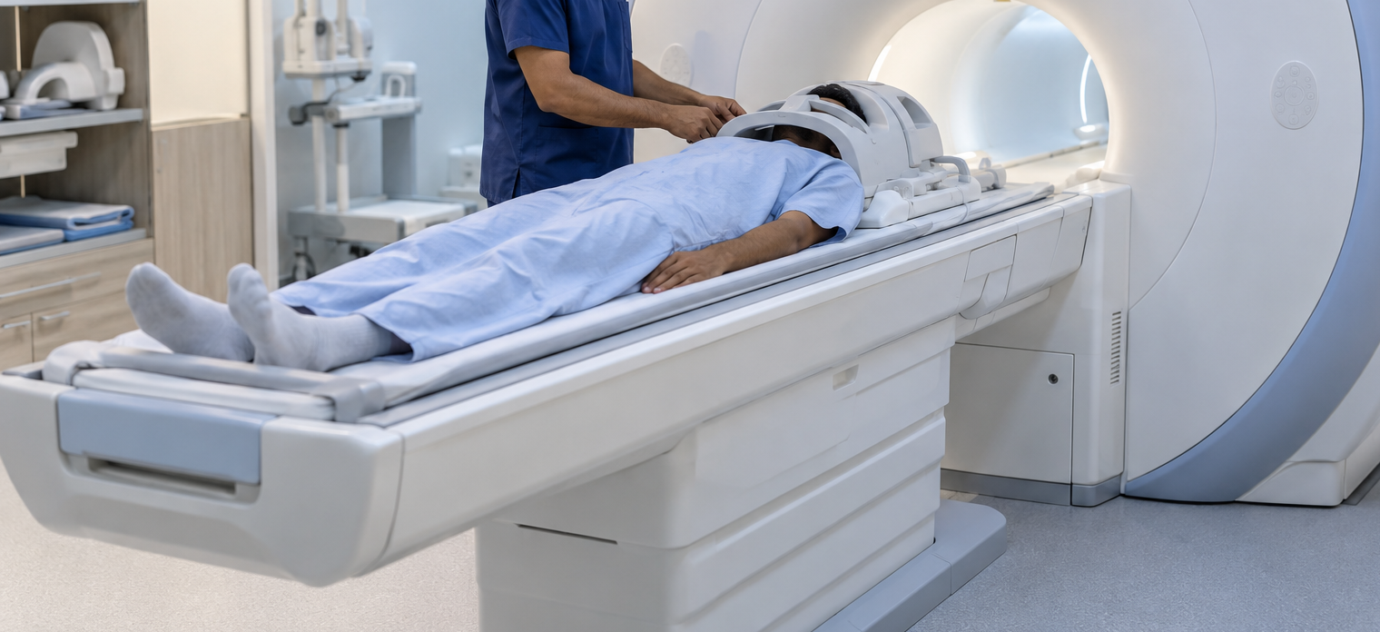
An MRI report containing terms such as disc bulge, degeneration, stenosis, nerve-root compression, or altered spinal cord signal can appear alarming. Yet an abnormal scan does not automatically mean that every finding is responsible for the patient's symptoms or that surgery is required. A spine MRI examination is most useful when imaging findings are interpreted together with the person's neurological history and physical examination.
For patients in Sakinaka and surrounding areas of Mumbai, Maharashtra, knowing when a spine MRI is appropriate can help avoid both unnecessary anxiety and delays in investigating meaningful neurological symptoms.
When Is Spine MRI Commonly Considered?
Many episodes of uncomplicated back pain improve without immediate advanced imaging. MRI becomes more relevant when symptoms or examination findings raise concern about nerve roots, the spinal cord, infection, tumors, significant injury, or another structural condition.
A doctor may consider MRI when there is:
- Persistent pain with neurological symptoms
- Progressive arm or leg weakness
- Numbness following a nerve distribution
- Significant walking or balance problems
- Suspected spinal cord compression
- Symptoms suggesting spinal infection or tumor
- Persistent radicular pain that has not improved as expected
- Relevant history requiring closer structural evaluation
The appropriate timing depends on the clinical situation.
Cervical, Lumbar and Thoracic MRI Examine Different Regions
The spine is divided into regions, and the scan should correspond to the suspected neurological location.
Cervical spine MRI examines the neck region and may be relevant when symptoms affect the arms, hands, legs, or spinal cord pathways.
Thoracic spine MRI evaluates the middle portion of the spine and may be required when symptoms suggest a lesion affecting this region.
Lumbar spine MRI is frequently used for lower-back symptoms associated with leg pain, numbness, weakness, or suspected nerve-root compression.
Scanning the correct area is more useful than ordering multiple regions without a clear clinical reason.
Why MRI Findings Need Careful Interpretation
Degenerative spinal changes become increasingly common with age. A scan may therefore identify disc bulges or other abnormalities even in someone who has few or no symptoms.
The critical question is whether the location and severity of an imaging abnormality match the patient's complaints and examination.
For example, a disc abnormality at one spinal level should not automatically be blamed for symptoms that follow an entirely different neurological distribution.
Neurological examination helps establish this correlation.
MRI Can Show More Than Disc Problems
Although spine MRI is commonly associated with slipped discs, it can provide information about many structures and conditions.
Depending on the region and imaging protocol, MRI may demonstrate:
- Disc degeneration or herniation
- Spinal canal narrowing
- Nerve-root compression
- Spinal cord compression
- Abnormal spinal cord signals
- Certain tumors
- Inflammatory lesions
- Infection-related changes
- Vertebral abnormalities
Contrast may be recommended in particular situations rather than for every routine spine scan.
When a Spine Problem Needs Faster Evaluation
Certain neurological symptoms should not be managed by simply scheduling routine imaging at a convenient future date.
New bladder or bowel dysfunction with neurological symptoms, numbness around the saddle region, rapidly progressive leg weakness, sudden major difficulty walking, or severe neurological changes following trauma require prompt medical assessment.
These symptoms can indicate significant compression or another neurological emergency.
Choosing Evaluation Before Choosing Treatment
An MRI result should guide care rather than dictate it automatically.
Some patients may need medicines, physiotherapy, rehabilitation, activity modification, or observation. Others with significant neurological compression may need evaluation by a spine surgeon or neurosurgeon. Inflammatory, infectious, neoplastic, or other neurological conditions require treatment directed at their underlying cause.
For patients in Sakinaka, Dr. Sachin Adukia can evaluate neurological symptoms, correlate them with spine imaging, and determine whether additional investigations or specialist referrals are appropriate.
The purpose of an MRI spine examination is not merely to find abnormalities. Its real clinical value lies in identifying structural changes that meaningfully correspond with the patient's symptoms, distinguishing incidental findings from significant disease, and helping guide the next stage of care.
Keywords