Headaches are extremely common and, by themselves, are usually not enough to indicate a brain tumor. The situation becomes different when headaches occur alongside new neurological changes or when a patient's symptoms develop an unusual and progressive pattern. Brain tumor diagnosis therefore depends on clinical assessment and appropriate imaging rather than assumptions based on a single symptom.
For patients and families in Bhandup, Mumbai, Maharashtra, knowing which changes deserve neurological attention can help separate routine symptoms from those requiring further investigation.
Headache Pattern Matters More Than Fear
Many people associate persistent headaches with brain tumors, but migraine, tension-type headache, sinus-related symptoms, medication overuse, sleep disruption, and several other conditions are much more common explanations.
During neurological evaluation, the doctor may ask whether the headache is new, whether its pattern has changed, how severe it is, and whether neurological symptoms accompany it.
Symptoms that can warrant further evaluation include:
- New seizures
- Persistent vomiting with neurological symptoms
- New weakness or numbness
- Changes in speech
- Unexplained visual disturbances
- Significant personality or cognitive changes
- Increasing balance difficulty
- Progressive coordination problems
These symptoms can have causes other than tumors, but they should not be dismissed without appropriate assessment.
The Neurological Examination Provides Direction
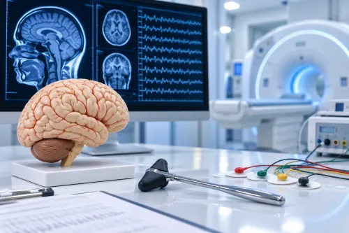
Before ordering tests, a neurologist typically evaluates different aspects of nervous-system function.
This may include checking eye movements, visual fields, facial movement, speech, muscle strength, sensation, reflexes, coordination, gait, and cognitive function.
The findings help identify whether symptoms could be arising from a particular part of the brain and whether imaging is indicated.
MRI and CT Serve Different Purposes
Brain imaging is central when a structural intracranial problem is suspected.
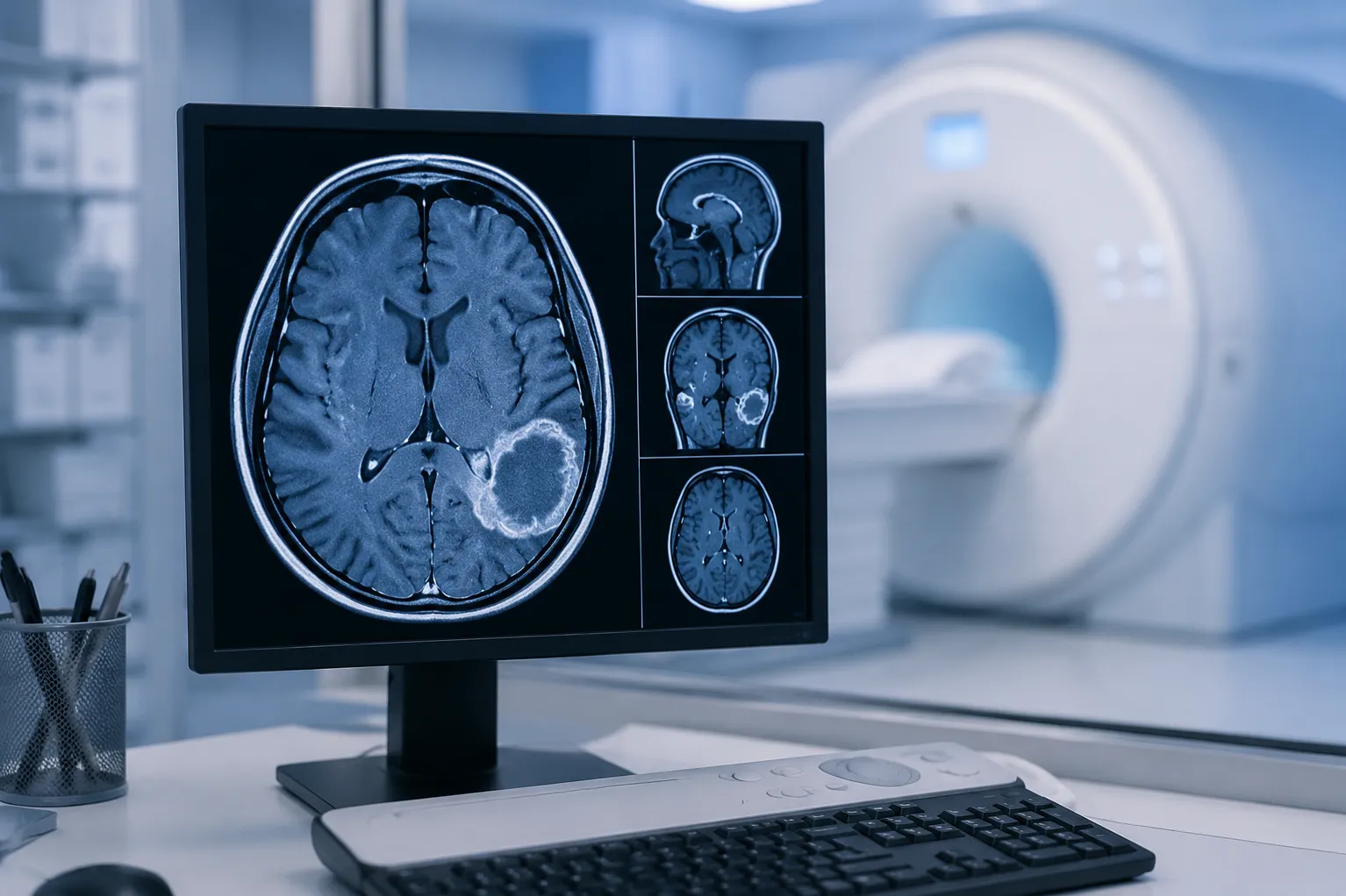
MRI provides detailed images of brain tissue and is frequently used when evaluating suspected tumors. Contrast-enhanced MRI may be recommended depending on the clinical situation.
CT scanning can also be useful, particularly in emergency situations or when rapid imaging is required. It may identify bleeding, significant swelling, hydrocephalus, calcification, or a mass.
Choosing an investigation depends on the symptoms, urgency, patient circumstances, and what clinicians need to determine.
Does a Scan Confirm Exactly What a Tumor Is?
Not always.
Imaging can reveal the location, approximate size, surrounding swelling, relationship to nearby structures, and characteristics of a lesion. These findings may suggest particular possibilities, but determining the exact tumor type can require tissue examination.
When appropriate, a neurosurgical team may consider biopsy or surgical removal. A pathologist then examines tissue to establish the histological diagnosis. Molecular testing is increasingly relevant for classifying several brain tumors and helping specialists plan subsequent management.
What Happens After an Abnormal Scan?
An abnormal MRI does not automatically mean immediate surgery.
Management depends on factors such as the type of lesion suspected, its location, size, growth pattern, symptoms, age and overall health of the patient.
The care pathway can involve neurology, neurosurgery, oncology, radiation oncology, radiology, and pathology.
Some lesions may require observation with follow-up imaging, while others need biopsy, surgery, radiotherapy, systemic treatment, or a combination of approaches.
Know When Symptoms Need Prompt Attention
A first seizure, sudden neurological deficit, severe rapidly developing headache, loss of consciousness, repeated vomiting with neurological deterioration, or new severe confusion needs urgent medical evaluation.
For patients in Bhandup, Dr. Sachin Adukia can assess neurological symptoms and determine when brain imaging or further specialist evaluation is appropriate.
Brain tumor diagnosis should be systematic rather than fear-driven. Recognizing concerning symptom combinations, performing a focused neurological examination, selecting appropriate imaging, and establishing the nature of an identified lesion provide a far more reliable pathway than trying to interpret headache symptoms alone.
Keywords